Posts

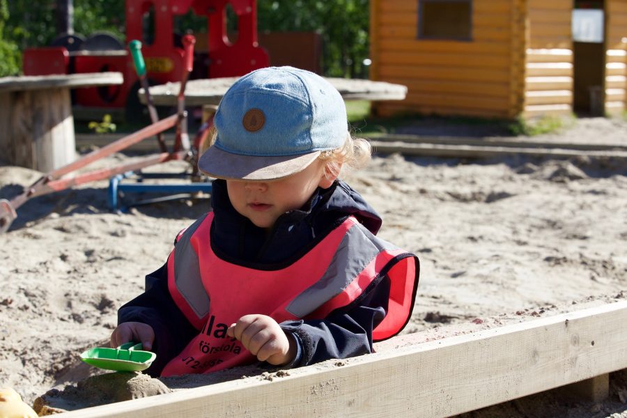
Shaping microbial exposures and the immune system in childhood: Can sandboxes be probiotic?

By Prof. Seppo Salminen, University of Turku, Finland

Gut microbiota researchers have established that microbial exposures in early life can be influential on health later in life. Children who develop asthma in early childhood, for example, have an altered gut microbiota linked with exposure to less diverse microorganisms in their first year. The ‘biodiversity hypothesis’ has been advanced recently, suggesting that western lifestyles and low biodiversity in urban environments reduce contact with microbes both via food and via the natural environment, presenting fewer opportunities for children to be exposed to a diversity of microbes in their earliest years and increasing the risk of non-communicable diseases. If this is the case, the environments of daycare and kindergarten facilities come under scrutiny as a source of microbial exposures at a crucial time of life. So is it beneficial to intervene in children’s environments to ensure more diverse microbial exposures? Can we enhance gut microbial diversity and richness in children through environmental interventions?

A new study provided proof that shaping children’s microbial exposures may be possible. The study was the first of its kind – a placebo-controlled, double-blinded study on the effect of environmental exposures on gut microbiota diversity and immune parameters in young children. The study used playground sandboxes at daycare facilities as sources of environmental microbial diversity and explored whether these could have effects on the children.

Six day-care centers in southern Finland were enrolled in the study, with two randomly assigned to intervention and four to placebo. Identical-looking playground sandboxes were used. Intervention sandboxes were filled with sand of glacial origin enriched with a known biodiversity powder (including commercial soil, deciduous leaf litter, peat, and Sphagnum moss; described in detail by Hui et al., 2019 ; Grönroos et al, 2018). In control centers the sand was regular sandbox sand and placebo peat material. Altogether, 26 children ages 3-5 participated in supervised play for 20 minutes in the morning and afternoon for two weeks. Researchers measured the composition of gut and skin microbiota, as well as blood immune markers.

The results demonstrated that exposure to diverse environmental microbiota enhanced both the bacterial richness and diversity of the skin bacterial community. The microbiome of the skin changed only in those children who had played in a sandbox enriched with natural materials. The authors also found that the daily exposure to higher microbial biodiversity resulted in positive differences in immune response. For instance, the authors reported shifts in skin microbiota associated with IL-10 and T cell frequencies. This provides the first evidence from a placebo-controlled, double-blinded study in young children showing the differential effects on microbiota and immunity of daily exposure to defined microbial biodiversity.

An interesting follow-up could be using sandboxes to deliver probiotics with a proven health impact to children. Since the sandbox microbes were shown to influence children’s immune systems, could researchers go one step further and modulate children’s microbiota in a targeted manner? A probiotic must be defined, shown to have a health benefit and administered in an efficacious dose. In the case of sandboxes, the health benefit would need to be demonstrated for a certain level or duration of environmental exposure.

Playgrounds and sandboxes require materials that tolerate heavy wear and tear and are safe at the same time. Such materials need to be kept free of unnecessary contamination as sandboxes, for example, can also be good reservoirs of some detrimental bacteria. Therefore, it could be important to have defined natural materials for a positive impact on health. In the future, we may see many creative approaches to ensuring children receive appropriate health-supporting microbial exposures early in life. However, creating probiotic approaches requires identification of specific microbes in the biodiversity powder.

Bifidobacteria in the infant gut use human milk oligosaccharides: how does this lead to health benefits?

By Martin Frederik Laursen, Technical University of Denmark, 2022 co-recipient of Glenn Gibson Early Career Research Prize

Breast milk is the ‘gold standard’ of infant nutrition, and recently scientists have zeroed in on human milk oligosaccharides (HMOs) as key components of human milk, which through specific interaction with bifidobacteria, may improve infant health. Clarifying mechanisms by which HMOs act in concert with bifidobacteria in the infant gut may lead to better nutritional products for infants.

Back in early 2016, I was in the middle of my PhD studies working on determinants of the infant gut microbiota composition in the Licht lab at the National Food Institute, Technical University of Denmark. I had been working with fecal samples from a Danish infant cohort study, called SKOT (Danish abbreviation for “Diet and well-being of young children”), investigating how the diet introduced in the complementary feeding period (as recorded by the researchers) influences the gut microbiota development 1,2. Around the same time, Henrik Munch Roager, PostDoc in the lab, was developing a liquid chromatography mass spectrometry (LC-MS)-based method for quantifying the aromatic amino acids (AAA) and their bacterially produced metabolites in fecal samples (the 3 AAAs and 16 derivatives thereof). These bacterially produced AAA metabolites were starting to receive attention because of their role in microbiota-host cross-talk and interaction with various receptors such as the Aryl Hydrocarbon Receptor (AhR) expressed in immune cells and important for controlling immune responses at mucosal surfaces 3,4. However, virtually nothing was known about bacterial metabolism of the AAAs in the gut in an early life context. Further, the fecal samples collected from the SKOT cohort were obtained in a period of life when infants are experiencing rapid dietary changes (e.g. cessation of breastfeeding and introduction of various new foods). Thus, we wondered whether the AAA metabolites would be affected by diet and whether these metabolites might contribute to the development of the infant’s immune system. Our initial results quickly guided us on the track of breastfeeding and bifidobacteria! Here is a summary of the story, published last year in Nature Microbiology5. (See the accompanying News & Views article here.)

We initially looked at the data from a subset of 59 infants, aged 9 months, from the SKOT cohort. Here we found that both the gut microbiome and the AAA metabolome were affected by breastfeeding status (breastfed versus weaned). It is well established that certain bifidobacteria dominate the bacterial gut community in breastfed infants due to their efficient utilization of HMOs – which are abundant components of human breastmilk 6. Our data showed the same, namely enrichment of Bifidobacterium in the breastfed infants, but also indicated that the abundance of specific AAA metabolites were dependent on breastfeeding.

Trying to connect the gut microbiome and AAA metabolome, we found striking correlations between the relative abundance of Bifidobacterium and specifically abundances of three aromatic amino acid catabolites – namely indolelactic acid (ILA), phenyllactic acid (PLA) and 4-hydroxyphenyllactic acid (4-OH-PLA), collectively aromatic lactic acids. These metabolites are formed in two enzymatic reactions (a transamination followed by a hydrogenation) of the aromatic amino acids tryptophan, phenylalanine and tyrosine. However, the genes involved in this pathway were not known for bifidobacteria. Digging deeper we discovered that not all Bifidobacterium species found in the infant’s gut correlated with these metabolites. This was only true for the Bifidobacterium species enriched in the breastfed infants (e.g. B. longum, B. bifidum and B. breve), but not post-weaning/adult type bifidobacteria such as B. adolescentis and B. catenulatum group.

We decided to go back to the lab and investigate these associations by culturing representative strains of the Bifidobacterium species found in the gut of these infants. Indeed, our results confirmed that Bifidobacterium species are able to produce aromatic lactic acids, and importantly that the ability to produce them was much stronger for the HMO-utilizing (e.g. B. longum, B. bifidum and B. breve) compared to the non-HMO utilizing bifidobacteria (e.g. B. adolescentis, B. animalis and B. catenulatum). Next, in a series of experiments we identified the genetic pathway in Bifidobacterium species responsible for production of the aromatic lactic acids and performed enzyme kinetic studies of the key enzyme, an aromatic lactate dehydrogenase (Aldh), catalyzing the last step of the conversion of aromatic amino acids into aromatic lactic acids. Thus, we were able to demonstrate the genetic and enzymatic basis for production of these metabolites in Bifidobacterium species.

To explore the temporal dynamics of Bifidobacteria and aromatic lactic acids and validate our findings in an early infancy context (a critical phase of immune system development), we recruited 25 infants (Copenhagen Infant Gut [CIG] cohort) from which we obtained feces from birth until six months of age. These data were instrumental for demonstrating the tight connection between specific Bifidobacterium species, HMO-utilization and production of aromatic lactic acids in the early infancy gut and further indicated that formula supplementation, pre-term delivery and antibiotics negatively influence the concentrations of these metabolites in early life.

Having established that HMO-utilizing Bifidobacterium species are key producers of aromatic lactic acids in the infant gut, we focused on the potential health implications of this. We were able to show that the capacity of early infancy feces to in vitro activate the AhR, depended on the abundance of aromatic lactic acid producing Bifidobacterium species and the concentrations of ILA (a known AhR agonist) in the fecal samples obtained from the CIG cohort. Further, using isolated human immune cells (ex vivo) we showed that ILA modulates cytokine responses in Th17 polarized cells – namely it increased IL-22 production in a dose and AhR-dependent manner. IL-22 is a cytokine important for protection of mucosal surfaces, e.g. it affects secretion of antimicrobial proteins, permeability and mucus production 7. Further, we tested ILA in LPS/INFγ induced monocytes (ex vivo), and found that ILA was able to decrease the production of the proinflammatory cytokine IL-12p70, in a manner dependent upon both AhR and the Hydroxycarboxylic Acid (HCA3) receptor, a receptor expressed in neutrophils, macrophages and monocytes and involved in mediation of anti-inflammatory processes 8,9. Overall, our data reveal potentially important ways in which bifidobacteria influence the infant’s developing immune system.

Figure 1 – HMO-utilizing Bifidobacterium species produce immuno-regulatory aromatic lactic acids in the infant gut.

Our study provided a novel link between HMO-utilizing Bifidobacterium species, production of aromatic lactic acids and immune-regulation in early life (Figure 1). This may explain previous observations that the relative abundance of bifidobacteria in the infant gut is inversely associated with development of asthma and allergic diseases 10–12 and our results, together with other recent findings13–15 are pointing towards aromatic lactic acids (especially ILA) as potentially important mediators of beneficial immune effects induced by HMO-utilizing Bifidobacterium species.


References

  1. Laursen, M. F. et al. Infant Gut Microbiota Development Is Driven by Transition to Family Foods Independent of Maternal Obesity. mSphere 1, e00069-15 (2016).
  2. Laursen, M. F., Bahl, M. I., Michaelsen, K. F. & Licht, T. R. First foods and gut microbes. Front. Microbiol. 8, (2017).
  3. Zelante, T. et al. Tryptophan catabolites from microbiota engage aryl hydrocarbon receptor and balance mucosal reactivity via interleukin-22. Immunity 39, 372–385 (2013).
  4. Sridharan, G. V. et al. Prediction and quantification of bioactive microbiota metabolites in the mouse gut. Nat. Commun. 5, 1–13 (2014).
  5. Laursen, M. F. et al. Bifidobacterium species associated with breastfeeding produce aromatic lactic acids in the infant gut. Nat. Microbiol. 6, 1367–1382 (2021).
  6. Sakanaka, M. et al. Varied pathways of infant gut-associated Bifidobacterium to assimilate human milk oligosaccharides: Prevalence of the gene set and its correlation with bifidobacteria-rich microbiota formation. Nutrients 12, 71 (2020).
  7. Keir, M. E., Yi, T., Lu, T. T. & Ghilardi, N. The role of IL-22 in intestinal health and disease. J. Exp. Med. 217, (2020).
  8. Peters, A. et al. Metabolites of lactic acid bacteria present in fermented foods are highly potent agonists of human hydroxycarboxylic acid receptor 3. PLoS Genet. 15, e1008145 (2019).
  9. Peters, A. et al. Hydroxycarboxylic acid receptor 3 and GPR84 – Two metabolite-sensing G protein-coupled receptors with opposing functions in innate immune cells. Pharmacol. Res. 176, (2022).
  10. Fujimura, K. E. et al. Neonatal gut microbiota associates with childhood multisensitized atopy and T cell differentiation. Nat. Med. 22, 1187–1191 (2016).
  11. Stokholm, J. et al. Maturation of the gut microbiome and risk of asthma in childhood. Nat. Commun. 9, 141 (2018).
  12. Seppo, A. E. et al. Infant gut microbiome is enriched with Bifidobacterium longum ssp. infantis in Old Order Mennonites with traditional farming lifestyle. Allergy Eur. J. Allergy Clin. Immunol. 76, 3489–3503 (2021).
  13. Meng, D. et al. Indole-3-lactic acid, a metabolite of tryptophan, secreted by Bifidobacterium longum subspecies infantis is anti-inflammatory in the immature intestine. Pediatr. Res. 88, 209–217 (2020).
  14. Ehrlich, A. M. et al. Indole-3-lactic acid associated with Bifidobacterium-dominated microbiota significantly decreases inflammation in intestinal epithelial cells. BMC Microbiol. 20, 357 (2020).
  15. Henrick, B. M. et al. Bifidobacteria-mediated immune system imprinting early in life. Cell 184, 3884-3898.e11 (2021).

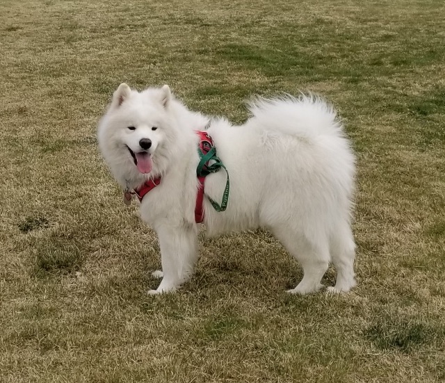
Children and dogs in a household share gut microbes – and these microbes are modified by a canine probiotic

From longtime family pets to ‘pandemic puppies’, dog ownership is seemingly more popular than ever. In households with children, scientists have found that a pet dog is one of the environmental factors that influences the gut microbiota in early life – but can the microbes that children and dogs share be modified?

A new study from ISAPP president Prof. Seppo Salminen (University of Turku, Finland) and colleagues recently explored the impact of a household dog on children’s gut microbiota, before and after the dogs were given a canine probiotic. Not only did the gut microbiota of dogs and children in the same household share features in common, but also the gut microbes of both shifted after dogs received a probiotic.

The study, which was part of a larger investigation, looked at families with at least one member who had allergic disease. Thirty-one of the families in the current study had dogs, and 18 families (the control group) did not. From each household, the fecal microbiota of one child (aged 5 or under) was tested. The fecal microbiota of the dogs was tested, and further, they received either a probiotic containing 3 canine-derived strains, or placebo.

The data supported previous observations that dogs and children share gut microbes: the children living with dogs had a distinct fecal microbiota composition. The most striking microbiota differences were a higher abundance of Bacteroides and short-chain fatty acid producing bacteria.

Moreover, when the household dogs were given a probiotic, both the dogs and the children living with them showed a gut microbiota shift, with a reduction in Bacteroides. (The exact probiotic strains were not tracked in the feces of either the dogs or the children.)

Were the changes beneficial? It’s not certain, since health outcomes in the children were not part of the study. But these findings provide more evidence for the effect of home environments and pets on the gut microbiota of children, and highlight the modifiability of both the dog’s and children’s gut microbiota. The ability to modify a child’s gut microbiota is of particular interest in the early years, when gut microbiota / immune interactions have the potential to shape health through the lifespan.

The study authors conclude, “Our promising data invite the idea that the compositional development of the gut microbiota in children is potentially modifiable by indirect changes in household pets and justify the further search of novel modes of intervention during critical period when the scene is set for the consolidation of the child later health.”

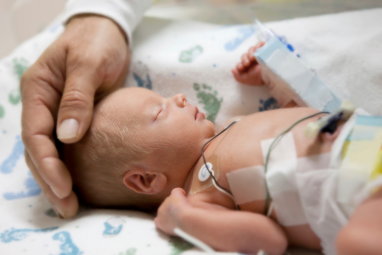
Probiotics to Prevent Necrotizing Enterocolitis: Moving to Evidence-Based Use

By Ravi Mangal Patel, MD, Msc, Associate Professor of Pediatrics, Emory University School of Medicine and Children’s Healthcare of Atlanta. rmpatel@nullemory.edu Twitter: @ravimpatelmd

Necrotizing enterocolitis (NEC) is one of the most lethal neonatal diseases, yet most people have never heard about it. The disease primarily affects preterm infants and is characterized by the development of intestinal inflammation. Clinically, the disease often manifests with an infant developing feeding intolerance or abnormal abdominal exam findings. The diagnosis is confirmed by abdominal x-ray or ultrasound. One of the key diagnostic radiographic findings is pneumatosis, which is air in the lumen of the bowel caused by gas-producing bacteria.

Dr Ravi Mangal Patel

NEC accounts for 1 out of every 10 deaths in US neonatal intensive care units. Among extremely preterm infants (those born at 22-28 weeks’ gestation) in the US, NEC is the most common single cause of death between 2 weeks and 2 months of age. Many infants with NEC undergo surgery to remove diseased bowel and those who recover and survive are at risk for long-term neurodevelopmental impairment and short bowel syndrome.

Decades of research into NEC have identified several key risk factors, including formula feeding, inconsistent feeding, abnormal intestinal oxygenation and [gut microbiota] dysbiosis. Studies have shown that dysbiosis, or abnormal intestinal colonization, is an important antecedent risk factor for the development of NEC. These studies have found that infants who develop NEC have an increase or bloom in the relative abundance of proteobacteria, compared to those who do not develop NEC. These proteobacteria, which contain a lipopolysaccharide coating, may lead to inflammation through their interaction with Toll-like receptor 4.

Given the role of dysbiosis in NEC, efforts to intervene by provision of probiotics to prevent NEC is a rational and extensively studied intervention, with over 63 randomized trials enrolling ~15,000 infants to date. The aforementioned meta-analysis, along with several others (Table 1), show probiotic supplementation results in large magnitude reductions in the risks of NEC and death and more modest reductions in the risks of late-onset sepsis. However, there is more limited data on extremely preterm infants and the quality or certainty of evidence for probiotics for the prevention of NEC was low in a recent Cochrane review.


Source: https://doi.org/10.1053/j.sempedsurg.2017.11.008

In the United States, an increasing number of centers have begun to routinely provide probiotics, with the greatest increase in use beginning in 2015. Observational studies evaluating routine probiotic use show benefits that are similar in magnitude to those from randomized trials, supporting the external validity of the results from the trials. This includes a large recent evaluation of probiotic use in the United States. Around the world, probiotic use is highly variable, from 100% of NICUs in New Zealand, 68% of NICUs in Germany, to 12% in the UK, 21% in Canada and 14% in the United States. Some of the variability in clinical use may be related to the uncertainty regarding the quality of commercially available probiotic products and need for clarity regarding strain-specificity of effects. There are many considerations both for and against routine use of probiotics to prevent NEC (Table 2). Current probiotic dietary supplements do not undergo FDA’s premarket review and approval requirements for safety and effectiveness or have to meet manufacturing and testing standards for drugs, and the potential risks were highlighted by a case of an infant death from a contaminated supplement. There is currently no FDA-approved live biotherapeutic product to prevent NEC.

Source: doi: 10.1016/j.earlhumdev.2019.05.009

Recent recommendations and guidance from ESPHGAN and the AGA also demonstrate that some medical organizations recognize the strength of the data in support of probiotic use to prevent NEC. It has been over two decades since the first study demonstrating the benefit of probiotic supplementation to prevent NEC in preterm infants. Now, more than ever, the evidence continues to accumulate regarding the beneficial effects of probiotic use in preterm infants as a compelling strategy to reduce the risks of both NEC and death. Therefore, considering the balance of potential risks and benefits including data from both randomized trials and routine implementation studies, my opinion is that the cumulative evidence to date supports routine probiotic use to prevent NEC and death in preterm infants.

As important is considering the parent voice regarding probiotic use. The NEC Society is a non-profit focused on NEC that has worked to incorporate the voice of the patient-family in clinical decisions.

Disclosures: Dr. Patel serves on the data-safety monitoring board of the Connection Study, which is a trial examining the use of an investigational probiotic to decrease the risk of NEC.

For further information, see this seminar by Dr. Patel: Practical Consideration for Probiotics in the NICU