Expert insights into skin microbiome research and translation


The microbiome of the body’s largest organ – the skin – captured popular attention in 2019 when a high-profile ad campaign for soap called it a “living layer” on the body. Although the gut microbiome has dominated scientific attention and funding for the past two decades, skin microbiome research is now gaining momentum as researchers uncover its important and unique contributions to health and disease.
ISAPP recently explored the latest developments in skin microbiome research through a series of four podcasts featuring conversations with leading researchers in the field. This article gives an overview of skin microbiome research and shares some of the most compelling insights from these conversations.
What is the skin microbiome?
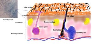
The skin microbiome includes all microorganisms residing on the skin: bacteria, fungi and archaea as well as viruses. The architecture of the skin includes three specialized layers: hypodermis, dermis, and epidermis. Microorganisms exist on the epidermis (outer layer) as well as in the hair follicles and glands. Major groups of microbes on the skin include Proteobacteria and Staphylococcus species, as well as commensal fungi such as Malassezia species.
Dr. Aayushi Uberoi PhD from Washington University in St. Louis (USA) notes that the skin microbiome varies across different locations of the body, with each area (whether dry, moist or oily) adapted to different physical demands and environmental exposures. These differences change the nutritional landscape for microorganisms in each area and affect microbial ecology, resulting in a skin microbiome that varies widely at different body sites. In general, healthy skin has a low pH, which is a key factor influencing the microorganisms that thrive there.
Studying the skin microbiome
Skin microbiome research has a set of unique challenges, as techniques used for sampling, sequencing and analysis must be appropriate for skin-associated microorganisms and tailored to the particular skin site (1). Sampling across a study must be consistent, with various levels of sampling invasiveness – from skin swabbing to tape-stripping to biopsy. Sampling challenges also arise from the fact that many areas of the skin are constantly picking up microorganisms from the external environment. Prof. Hariom Yadav PhD, from the University of South Florida (USA) says human skin has a stable (or core) microbiome, plus a transient microbiome that depends on recent exposures (2). Depending on the research question, this may require longitudinal or time series sampling of a skin site.
Regardless of sampling techniques, the low microbial biomass of skin samples (compared, for example, with gut samples), makes them susceptible to contamination. Negative and kit controls are critical in skin microbiome analysis to account for contaminants and observe the signals of interest.
Uncovering skin microbiome functions
A growing number of studies show the skin microbiome’s metabolic activities affect host cells to continually maintain the health of the skin. The microorganisms are required for essential skin functions, such as maintaining the epidermal barrier to protect against pathogens and dehydration, notes Dr. Uberoi. It’s known that the aryl hydrocarbon receptor (AhR) (3), a transcription factor (protein) expressed in every skin cell type, controls inflammation and protects against skin barrier damage and infection, making it the target of several effective skin therapeutics, including coal tar and tapinarof (a topical therapeutic) (4). Dr. Uberoi’s work has found that AhR can be activated by tryptophan metabolites originating from skin microbes, thereby improving skin barrier repair and function (5).
Discovering how skin microorganisms contribute to skin disease
One important area of discovery has been the contributions of skin microorganisms to a range of dermal diseases, including atopic dermatitis, psoriasis, acne, rosacea and wound-associated infections.
The research of Dr. Maria Teresa García-Romero, MD MPH from the National Institute of Pediatrics in Mexico City focuses on the role of skin microorganisms in atopic dermatitis (sometimes known as eczema). This condition can have profound effects on quality of life for affected children and their families, as well as affected adults, yet treatment options are limited.
Dr. García-Romero explains the growing evidence that the skin-dwelling bacterial species Staphylococcus aureus plays a role in initiating atopic dermatitis. Human studies show that when atopic dermatitis arises, S. aureus increases in abundance on the skin while the overall skin microbiome diversity decreases (6). Levels of S. aureus also correlate with severity of symptoms. Mechanistic research has built on this idea and shown that S. aureus thrives when the host decreases its production of antimicrobial peptides on the skin, compromising the epidermal barrier (7). Dr. García-Romero describes a cycle of inflammation involving S. aureus: the bacteria are proinflammatory and use proteases to drive skin inflammation, and in turn they benefit from an inflammatory environment to thrive on the skin. This results in symptoms of atopic dermatitis.
Further underlining the connection of S. aureus to atopic dermatitis is the fact that known effective treatments, such as antibiotics, are shown to reduce this species of bacteria on the skin (8). However, S. aureus can counter the effect of these treatments: a systematic review and meta-analysis from Dr. García-Romero’s group found S. aureus isolates from people with atopic dermatitis had less than optimal susceptibility to commonly used antimicrobials, especially in lower middle-income and upper middle-income countries (9).
Leveraging what’s known about S. aureus and atopic dermatitis may lead to more therapeutic options in the years ahead. Commercial development underway includes novel probiotic strains as topicals or live biotherapeutics to subdue virulent S. aureus on the skin in atopic dermatitis. A number of ingested probiotics, backed by clinical data, are already commercially available for atopic dermatitis.
Uncovering how skin bacteria drive other diseases
Skin microbes, however, have effects that go far beyond the skin itself. Research has found that microorganisms inhabiting the skin elicit unique immune responses with systemic effects. Dr. Nathan Archer PhD from Johns Hopkins Medicine (USA) describes how the skin microorganisms can shape immune reactions that cause inflammation even in the lungs. His lab is interested in understanding the connection between skin microorganisms and the ‘atopic march’ (a progression of allergic diseases, including atopic dermatitis, that can occur through infancy and childhood).
Dr. Archer’s lab uses a range of in vivo preclinical models, including whole animal optical imaging (10) to track bacterial clearance and host immune responses, human skin organotypic culture models and novel humanized mouse models with both human skin and immune cells. Using these tools, he and colleagues found that S. aureus on the skin can become pathogenic when it starts producing IL‑36 (pro-inflammatory small proteins) (11), intensifying allergic reactions on the skin as well as in the lungs. This may be a key mechanism in hard-to-treat neutrophilic asthma.
As a strategy for preventing skin and allergic diseases, Dr. Archer’s group is exploring a vaccine for S. aureus. However, so far any vaccines specifically targeting this bacterium have not succeeded because S. aureus has so many methods for overcoming human immune responses. A relevant multivalent vaccine targeting immune-active proteins is currently being developed and is in Phase 1 clinical trials.
Exploring microbiome-targeted interventions for skin aging
Apart from skin disease, skin aging is another key topic of interest in the skin microbiome field. The skin microbiome constantly changes through the lifespan, with increased age altering skin surface lipids and pH to favor a reduction in Propionibacteria and an increase in Proteobacteria. Acinetobacter is particularly increased in those with premature skin aging (12).
Skin microbiome interventions (probiotics and postbiotics) (13) are a promising way to address the cosmetic effects of skin aging as well as aging-related disease. However, Prof. Yadav says that because of the large skin microbiome differences from person to person and in specific age groups, new technologies or products may have to be designed in a personalized manner and may not be applicable to everyone. Prof. Yadav’s lab conducted experiments with lactobacilli and found strain-dependent effects of probiotics on skin anti-aging; for some strains, these effects persisted when the inactivated bacteria (postbiotics) were used (14). Both topical and ingested probiotics and other biotics show promise for this use.
Shaping the future of skin microbiome research
Skin microbiome research is gaining momentum as more links to skin health and whole-body health are uncovered. Efforts to develop specialized techniques for studying the skin microbiome are proving worthwhile, as new biotic-based approaches to maintaining skin health begin to emerge. Realizing this potential will depend on continued collaboration across multiple disciplines, including microbiology, dermatology, immunology, and other fields, and on a commitment to standardization and innovative techniques that enable further understanding of this visible and important organ of the body.
References:
(1) Grogan MD, Bartow-McKenney C, Flowers L, Knight SAB, Uberoi A, Grice EA. Research Techniques Made Simple: Profiling the Skin Microbiota. J Invest Dermatol. 2019 Apr;139(4):747-752.e1. doi: 10.1016/j.jid.2019.01.024.
(2) Pistone D, Meroni G, Panelli S, D’Auria E, Acunzo M, Pasala AR, Zuccotti GV, Bandi C, Drago L. A Journey on the Skin Microbiome: Pitfalls and Opportunities. Int J Mol Sci. 2021 Sep 12;22(18):9846. doi: 10.3390/ijms22189846.
(3) Uberoi A, Bartow-McKenney C, Zheng Q, Flowers L, Campbell A, Knight SAB, Chan N, Wei M, Lovins V, Bugayev J, Horwinski J, Bradley C, Meyer J, Crumrine D, Sutter CH, Elias P, Mauldin E, Sutter TR, Grice EA. Commensal microbiota regulates skin barrier function and repair via signaling through the aryl hydrocarbon receptor. Cell Host Microbe. 2021 Aug 11;29(8):1235-1248.e8. doi: 10.1016/j.chom.2021.05.011. Epub 2021 Jul 1.
(4) van den Bogaard EH, Esser C, Perdew GH. The aryl hydrocarbon receptor at the forefront of host-microbe interactions in the skin: A perspective on current knowledge gaps and directions for future research and therapeutic applications. Exp Dermatol. 2021 Oct;30(10):1477-1483. doi: 10.1111/exd.14409. Epub 2021 Jul 8.
(5) Uberoi A, Bartow-McKenney C, Zheng Q, Flowers L, Campbell A, Knight SAB, Chan N, Wei M, Lovins V, Bugayev J, Horwinski J, Bradley C, Meyer J, Crumrine D, Sutter CH, Elias P, Mauldin E, Sutter TR, Grice EA. Commensal microbiota regulates skin barrier function and repair via signaling through the aryl hydrocarbon receptor. Cell Host Microbe. 2021 Aug 11;29(8):1235-1248.e8. doi: 10.1016/j.chom.2021.05.011. Epub 2021 Jul 1.
(6) Kim J, Kim BE, Ahn K, Leung DYM. Interactions Between Atopic Dermatitis and Staphylococcus aureus Infection: Clinical Implications. Allergy Asthma Immunol Res. 2019 Sep;11(5):593-603. doi: 10.4168/aair.2019.11.5.593.
(7) Nakatsuji T, Chen TH, Two AM, Chun KA, Narala S, Geha RS, Hata TR, Gallo RL. Staphylococcus aureus Exploits Epidermal Barrier Defects in Atopic Dermatitis to Trigger Cytokine Expression. J Invest Dermatol. 2016 Nov;136(11):2192-2200. doi: 10.1016/j.jid.2016.05.127. Epub 2016 Jul 2.
(8) Khadka VD, Key FM, Romo-González C, Martínez-Gayosso A, Campos-Cabrera BL, Gerónimo-Gallegos A, Lynn TC, Durán-McKinster C, Coria-Jiménez R, Lieberman TD, García-Romero MT. The Skin Microbiome of Patients With Atopic Dermatitis Normalizes Gradually During Treatment. Front Cell Infect Microbiol. 2021 Sep 24;11:720674. doi: 10.3389/fcimb.2021.720674.
(9) Elizalde-Jiménez IG, Ruiz-Hernández FG, Carmona-Cruz SA, Pastrana-Arellano E, Aquino-Andrade A, Romo-González C, Arias-de la Garza E, Álvarez-Villalobos NA, García-Romero MT. Global Antimicrobial Susceptibility Patterns of Staphylococcus aureus in Atopic Dermatitis: A Systematic Review and Meta-Analysis. JAMA Dermatol. 2024 Nov 1;160(11):1171-1181. doi: 10.1001/jamadermatol.2024.3360.
(10) Archer NK, Wang Y, Ortines RV, Liu H, Nolan SJ, Liu Q, Alphonse MP, Dikeman DA, Mazhar M, Miller RJ, Anderson LS, Francis KP, Simon SI, Miller LS. Preclinical Models and Methodologies for Monitoring Staphylococcus aureus Infections Using Noninvasive Optical Imaging. Methods Mol Biol. 2020;2069:197-228. doi: 10.1007/978-1-4939-9849-4_15.
(11) Patrick GJ, Liu H, Alphonse MP, Dikeman DA, Youn C, Otterson JC, Wang Y, Ravipati A, Mazhar M, Denny G, Ortines RV, Zhang E, Miller RJ, Dillen CA, Liu Q, Nolan SJ, Nguyen K, Marcello L, Do DC, Wier EM, Zhang Y, Caviness G, Klimowicz AC, Mierz DV, Fine JS, Sun G, Goldbach-Mansky R, Marusina AI, Merleev AA, Maverakis E, Garza LA, Milner JD, Gao P, Ramanujam M, Raymond EL, Archer NK, Miller LS. Epicutaneous Staphylococcus aureus induces IL-36 to enhance IgE production and ensuing allergic disease. J Clin Invest. 2021 Mar 1;131(5):e143334. doi: 10.1172/JCI143334.
(12) Xu YN, Pu M, Raut J, Du Y, Qiu Q, Hermanson K, Xu Y, Doraiswamy C, Chu CC. Skin microbiome as a signature of premature ageing: enhancement with a retinyl propionate-containing topical product. Br J Dermatol. 2025 Oct 21;193(Suppl 2):ii24-ii31. doi: 10.1093/bjd/ljaf098.
(13) Prajapati SK, Lekkala L, Yadav D, Jain S, Yadav H. Microbiome and Postbiotics in Skin Health. Biomedicines. 2025 Mar 25;13(4):791. doi: 10.3390/biomedicines13040791.
(14) Challa V, Prajapati SK, Gangani S, Yadav D, Lekkala L, Jain S, Yadav H. Microbiome-Aging-Wrinkles Axis of Skin: Molecular Insights and Microbial Interventions. Int J Mol Sci. 2025 Oct 15;26(20):10022. doi: 10.3390/ijms262010022. PMID: 41155313; PMCID: PMC12564825.